Technology
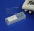
In order to gain insight into gene functions and changes in gene expression in plant organisms we work with cutting edge microarray technology. Microarrays (DNA chips, µ-arrays) are solid support substrates (mostly glass and plastic), coated with a reactive surface that binds DNA fragments. These are then processed by special equipment outlined below.
Microarrays
We spot cDNA or synthetic oligonucleotides onto glass slides using our contact or non-contact spotters in a density with up to 3000 spots per square centimetre. Thus one slide can represent up to 25000 single DNA fragments, originating from different genes.
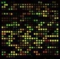
Microarrays can be used for hybridization experiments and work like classical dot-blot experiments, where one dot represents DNA fragment (one EST).
Liquid Handling
Material from our cooperation partners is transferred as bacterial colonies either in glycerol stock or on solid medium. According to the QC workflow, plasmid DNA is extracted from every single bacterial colony, and the insert is amplified by PCR reaction. After quality control of the PCR product, aliquots of the PCR reaction are stored at -20°C.
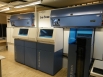
PCR & Gel Documentation
PCR-fragments, the raw material of cDNA arrays are amplified in PCR machines (MJ Dyade) and checked on agarose gels. The gels are analysed and documented by a hard- and software package (GelMaster®) that has been especially designed for high sample numbers and automated fragment analysis without manual interaction. The analysed data (Picture/size/amount/quality) are automatically stored in the PICME Database.
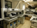
Spotter
Our institute is equipped with two different spotters, relying on different technologies. The machine used mainly for low density chips in surface development is a piezo-electric non-contact spotter. For high density chips, a contact spotter with up to 48 pins from Genemashines (Omnigrid) is used. This machine is capable of spotting up to 24.000 spots (DNA fragments), when spot-to-spot distance is 260µm. The spot diameter depends on the buffers used for spotting.
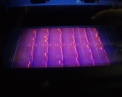
Reader
In order to analyse the DNA fragments of two different plants simultaneously, we use two different fluorescent dyes (Cy3, Cy5) to label the different individuals. We then spot both onto the same chip and analyse them in one single experiment. Chip readout (analysis of the hybridization experiment) is performed by a special fluorescent scanner (We use the Genepix scanner). This scanner renders a multicolour picture of the chip-surface for analysis.
Storage
For reliable management, individual samples in up to 4 replicate copies are stored within a modular -20°C/-80°C automated storage unit with an initial capacity of over 8.000 microtiter plates. With this robotic solution, our sample management (storage, replication and distribution of material upon request) is at the forefront of Biosample handling.